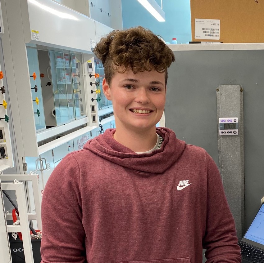
Trainees Appointed in 2024
-
Adriana Sordida
Harnessing Protein Language Models to Identify Novel Sterol and Steroid-Binding Proteins: Advancing Physiology and Functional Insights
Steroids are essential lipids that play a critical role in maintaining cellular membrane integrity and regulating hormonal and metabolic processes, yet many of the proteins that bind to these molecules remain unidentified or uncharacterized. My research leverages recent advances in machine learning, particularly Protein Language Models (PLMs), to predict and identify novel proteins that interact with sterols and steroids. A significant focus is on discovering bile acid-interacting proteins in both humans and gut microbes, as bile acids, which are co-metabolites produced by the host and modified by gut microbiota, are involved in crucial signaling pathways. Recent findings suggest that these novel bile acids may interact with unidentified proteins, and by applying PLM-based methods, I aim to uncover proteins that mediate these interactions, offering new insights into host-microbiome communication. By integrating PLM and machine learning tools, this research seeks to discover novel proteins that could have significant implications for both human physiology and our understanding of sterol-related functions across different organisms.
-
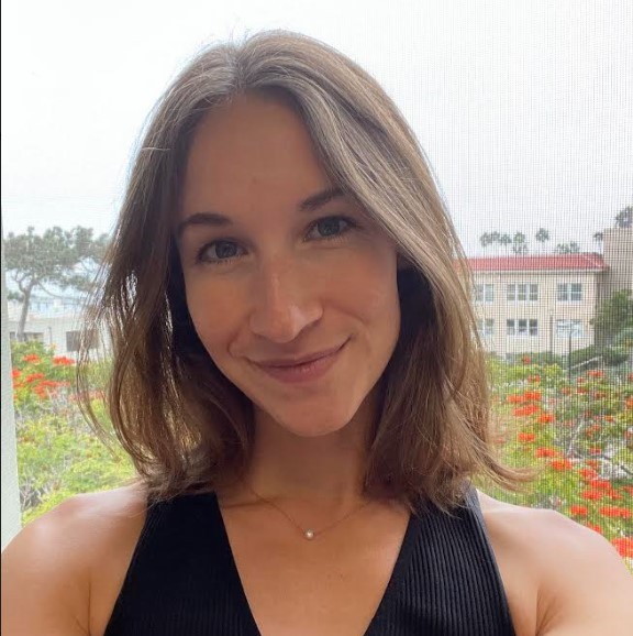
Alexa Perez
Structural and biochemical investigation of ncRNA and RNA binding protein interactions
Focus on the structural interactions between novel non-coding RNAs and the formation of RNP with predicted RNA binding proteins. Structural information will be used to aid the biochemical evaluation of RNA and protein functionality.
-
Anna Skrip
Uncovering the evolutionary history and medicinal potential of single-component flavin-dependent halogenases: an integrated approach
We aim to harness the power of selectivity and green chemistry with flavin-dependent halogenases. We utilize bioinformatics and enzyme characterization to understand nature’s evolved mechanistic preferences and inform novel biosynthetic approaches in drug development.
-
Brian Pham
Structural and functional characterization of RNA polymerase pausing and riboswitch co-transcriptional folding
My research focuses on how RNA polymerase pausing influences the co-transcriptional folding and function of riboswitches, regulatory elements in mRNAs that directly bind ligands to modulate gene expression. Specifically, I aim to uncover the mechanisms that link riboswitch folding kinetics to their regulatory activity.
-
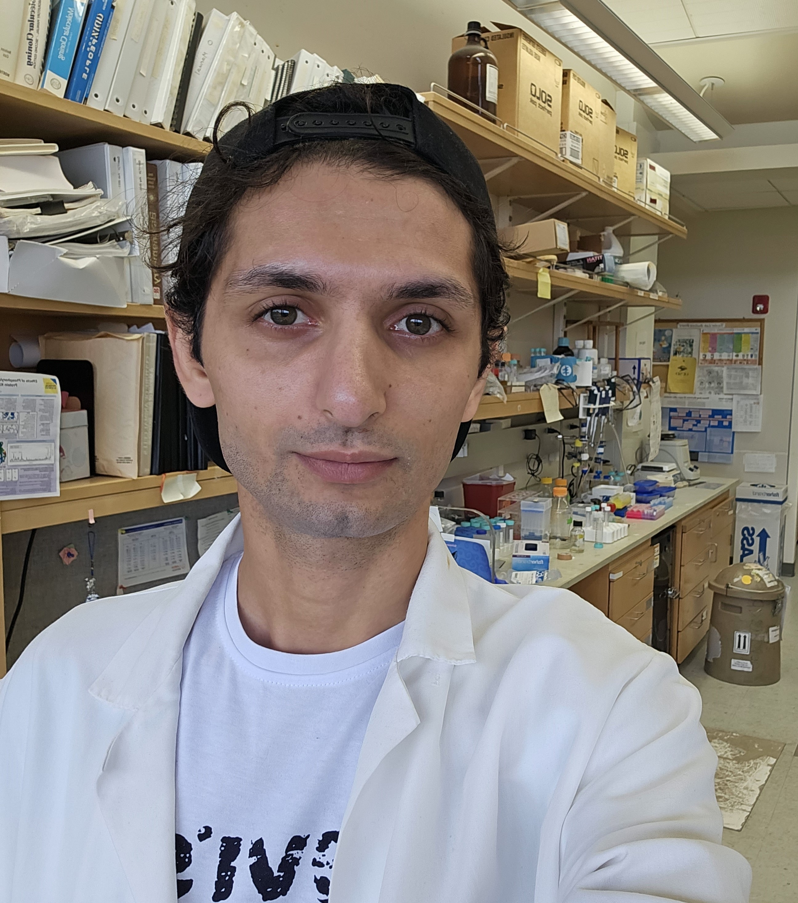
Caesar Tawfeeq
Conformational Dynamics of Wildtype and R201C Mutant G Stimulatory Protein Isoforms
As part of the Sunahara lab, my research involves applying computational biophysics tools and experimental techniques to characterize the conformational dynamics and molecular mechanisms of heterotrimeric G proteins. I am currently interested in understanding the differences between wildtype and R201C mutant of both the long and short splicing isoforms of G stimulatory proteins.
-
Christine Vu
Elucidating ERCC6L2-Mediated Liquid-Liquid Phase Separation and Protein Interactions in DNA Repair Pathways
This study investigates the role of ERCC6L2 in DNA repair, focusing on its potential involvement in liquid-liquid phase separation (LLPS) to form biological condensates. We aim to elucidate the dynamics of ERCC6L2 LLPS and its protein-protein interactions using live cell imaging and TurboID methodology. Findings will provide insights into the molecular mechanisms of DNA repair, highlighting ERCC6L2’s function in damage recognition and pathway selection, with implications for therapeutic targeting in DNA repair-related conditions.
-
Emerson Hall
Investigating high mannose binding lectins for applications as potential antiviral molecules
I aim to characterize the glycotypes of three unique lectins Oscillatoria agardhii agglutinin (OAA), Cyanovirin-N (CVN), and Griffithsin (GRFT) using biophysical techniques. Biolayer interferometry (BLI), nuclear magnetic resonance (NMR), isothermal titration calorimetry (ITC) will be utilized to provide kinetic, structural, and thermodynamic information respectively. I hope to dissect the factors underlying lectin specificity and mechanism behind multivalency.
-
Jaqueline Lanzalotto
Mechanisms of RNA Localization in Aspergillus Nidulans
Protein synthesis is an essential component of cellular growth, survival and maintenance. Newly synthesized proteins can be transported by the intracellular trafficking machinery to where they are required in the cell. However, proteins can be locally translated rather than trafficked from their site of synthesis to their final destination. mRNA transport plays a pivotal role in enabling precise, on-site translation of transcripts. My research aims to understand the molecular mechanisms of mRNA localization in the filamentous fungus Aspergillus nidulans.
-
Kezia Jemison
Title: Probing Receptor Binding Domain Dynamics of the SARS-CoV-2 Spike Protein
I investigate the dynamics of the SARS-CoV-2 spike protein’s Receptor Binding Domain (RBD) within the Wuhan-2019/Wild-type (WT), Delta (Δ), and Omicron (Ο) strains via Hydroxyl Radical Protein Footprinting for comparison to Amaro Lab predictions aiming to explain the observed increased COVID-infectivity of emerging variants.
Trainees Appointed in 2023 and reappointed in 2024
-
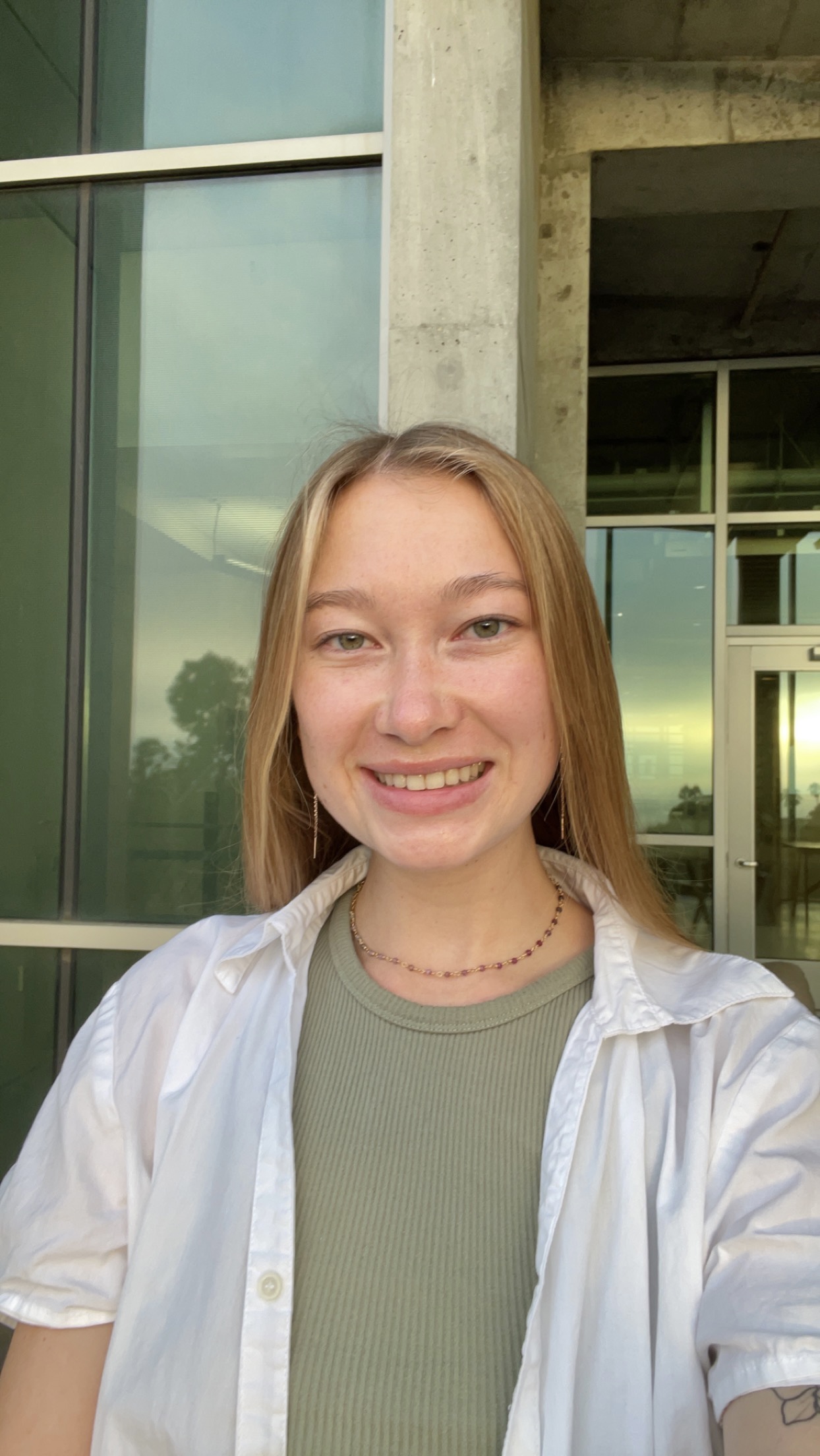
Kelly Isbell
Email: kisbell@ucsd.edu
Undergrad Institution: University of Virginia
Current Program at UC San Diego: Biochemistry and Molecular Biophysics
Advisor: Itay Budin
Mechanisms of PTCH1-Mediated Membrane Remodeling in Hedgehog SignalingThe hedgehog (Hh) pathway receptor Patched (Ptch1) suppresses the G-protein coupled receptor Smoothened (Smo). Ligands that inhibit Ptch1, such as sonic hedgehog in vertebrates, control tissue patterning during embryogenesis and tissue repair post-embryogenesis. Insufficient Hh signaling leads to developmental defects, whereas overactivation leads to ectodermal cancers including basal cell carcinoma (BCC) and medulloblastoma. Loss-of-function Ptch1 mutations are responsible for 85% of BCCs, a cancer for which more than 1 million US patients are treated for each year. Despite the narrow equilibrium necessary for proper Hedgehog signaling, exactly how Ptch1 regulates Smo is unknown. Ptch1 has been proposed to transport cholesterol across or out of the plasma membrane (PM), which goes hand-in-hand with findings that Smo is itself activated by cholesterol. The ability of cholesterol to act as a second messenger is surprising due to its high abundance in the PM and ability to rapidly flip-flop between bilayer leaflets. Sequestering interactions between sphingomyelin (SM) and cholesterol have been proposed to limit the accessibility of cholesterol to protein binding partners, but have not been rigorously tested. Using FRET and NMR-based assays, I aim to biophysically characterize the effects of SM sequestration on cholesterol chemical activity and interleaflet flip-flop kinetics. Then, utilizing Ptch1 proteoliposomes, I will elucidate the means and direction through which Ptch1 remodels membrane cholesterol as well as the conformational dynamics that dictate Ptch1 activity and inhibition. Finally, orchestrating a powerful combination of confocal microscopy, solvatochromatic dyes, engineered protein biosensors, and pharmacological and genetic manipulations, I will investigate the fundamental interactions between Ptch1, Smo, SM, and cholesterol that dictate Hh signaling in biophysically complex, living cell membranes. My ultimate goal is to generate a seamless model that describes how Ptch1-mediated remodeling of membrane cholesterol accessibility or activity on other lipid species regulates Smo activation.
-
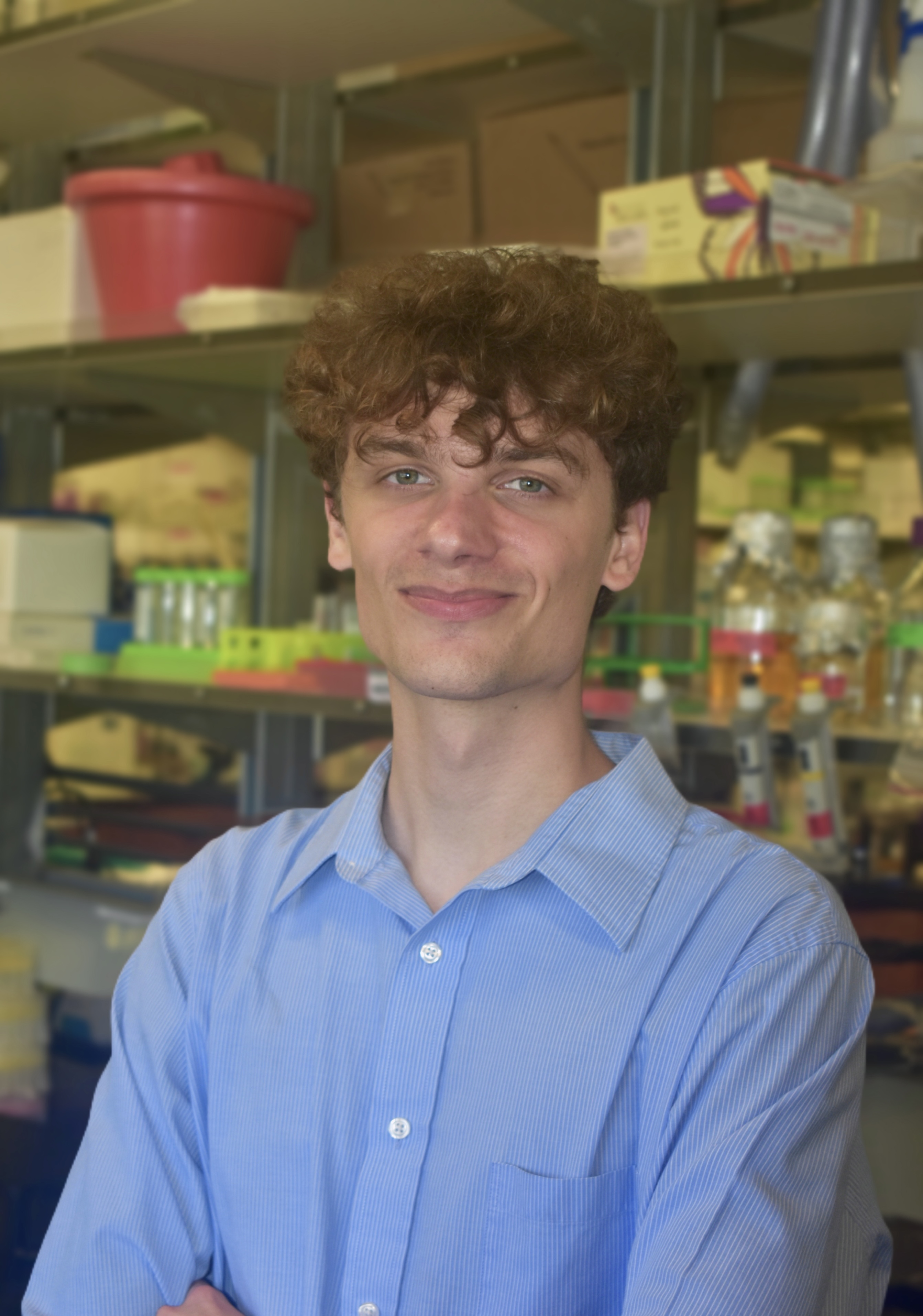
Benjamin Pollak
Email: brpollak@ucsd.eduUndergraduate institution: Boston UniversityCurrent program at UCSD: Biochemistry and Molecular BiophysicsAdvisor: Alexis KomorDevelopment of a mammalian cell directed evolution platform for base editors using TadATherapeutic genome editing can in theory cure genetic diseases by correcting underlying mutations at the genetic level. However, traditional genome editing methods that use Cas9-induced double-strand breaks (DSBs) suffer from low efficiencies and precision and are therefore not therapeutically viable for the correction of single nucleotide polymorphisms. Addressing this need, my advisor Alexis Komor pioneered DNA base editing, an innovative technology that enables accurate, precise, and programmable modifications of individual DNA bases that avoids the use of DSBs. I intend to develop and optimize a mammalian cell directed evolution platform for base editing, generate a large dataset of functional mutations in a successful DNA base editing enzyme (TadA), use in silico modeling to functionally characterize their roles in structure and DNA editing catalysis, and apply this system to other plausible DNA editing proteins to unlock additional classes of base editors. -
Alex Plonski
Email: aplonski@ucsd.edu
Undergrad Institution: University of Colorado, Denver
Current Program at UC San Diego: Biochemistry and Molecular Biophysics
Advisor: Galia Debelouchina
Structural and Mechanistic Investigations of Calcification in Age-Related Macular DegenerationAge-related macular degeneration (AMD) is a progressive eye disease that is most prevalent within aging populations and can lead to significant vision loss. AMD affects the macula on the central retina, where pathological extracellular deposits called drusen can be found between the sub-retinal pigment epithelium and Bruch’s membrane. These deposits consist of a core made up of extracellular lipids enriched with cholesterol, which are encased by calcium-phosphate debris known as hydroxyapatite with proteins associated on the surface. The protein substrates include the blood protein Vitronectin, and Alzheimer’s Disease associated proteins Amyloid beta and Tau. Despite the significance of drusen and their association with both AMD and neurodegenerative diseases, the understanding of their molecular-level structure, mechanisms of assembly, and the role of the associated proteins remains limited. Gaining insights into these aspects will be crucial in understanding the role of drusen and calcification in AMD progression and may expose opportunities for diagnostic and therapeutic interventions that will have a significant impact on public health, especially in the aging populations disproportionately affected by AMD. Therefore, I am employing a range of biophysical techniques, such as fluorescence microscopy, aggregation assays, and solid-state nuclear magnetic resonance methods to investigate the structural, chemical, and physical properties of these calcified deposits. I am aiming to elucidate the mechanism of assembly of drusen, as well as structurally investigate the protein-mineral interactions within drusen.
-
Evan Wen
Email: evwen@ucsd.eduUndergrad institution: HKUSTCurrent program at UC San Diego: Biochemistry and Molecular BiophysicsAdvisor: Dr. Jin ZhangMechanisms of compartmentalized GPCR-mediated ERK signalingThe aim of my research is to elucidate the spatiotemporal activity network and molecular mechanisms of compartmentalized GPCR-mediated ERK signaling. -
Suzanne Enos
Molecular insights into Asp/Glu transport by the Mitochondrial Carrier Family
In humans, tight regulation of aspartate and glutamate concentrations in the mitochondria and cytosol is maintained by the aspartate/glutamate carriers (AGCs) of the inner mitochondrial membrane, also known as SLC25A12 (AGC1/aralar) and SLC25A13 (AGC2/citrin) – regulation that is critical for the citric acid cycle, gluconeogenesis, myelin synthesis in neurons, and neurotransmission. Through structural characterization by cryogenic electron microscopy (cryoEM) and complementary biochemical study, I aim to elucidate the transport mechanism, substrate specificity, and regulation of AGC1 and AGC2.
-
Lydia Chambers
Email: lychambers@ucsd.edu
Undergrad Institution: Gonzaga University
Current Program at UC San Diego: Biochemistry and Biophysics
Advisor: Kevin Corbett and Elizabeth KomivesBacterial antiviral defense pathways encode eukaryotic-like ubiquitination systems
"Ubiquitin (Ub) is a highly conserved protein involved in the regulation of a cell. It is attached to a target proteins through the process of ubiquitination, which typically leads to the target’s degradation. Ubiquitination requires the coordinated function of an enzymatic cascade utilizing a Ub-activating enzyme (E1), a Ub-conjugating enzyme (E2), and a Ub-protein ligase (E3). In some systems, Ub will be substituted for a Ubiquitin-like protein (Ubl) which operates as Ub in the cascade but has a unique biological function. I am studying the recently identified BilABCD ubiquitination systems, which are present in bacteria and contain four proteins: a Ubl, CEHH (an E2 protein), a canonical E1 protein, and a deubiquitinase (DUB) which has been identified as a JAB/JAMM family peptidase. These bacterial defense systems protect the host from bacterial viruses known as bacteriophages (phage). The goal of my research is to characterize these systems by determining their target, the structures of all four proteins, and their biological role. I have determined two structures of the type II BilABCD E1-E2-Ubl complex. The first structure shows the E1 CYS domain in a downward conformation, positioning the E1 catalytic cysteine in the E1 adenylation site (Fig 1a). The second conformation shows the CYS domain flipped upward, now mimicking the transfer of Ubl from E1 to the catalytic cysteine on E2 (Fig 1b). When these structures are compared to canonical E1-E2-Ub/Ubl complexes, significant structural homology between the bacterial and eukaryotic systems is demonstrated (Fig 1c). I have also confirmed that the BilABCD systems are fully active Ubl conjugation systems and mutation of the E1 (C417) and E2 (C138) catalytic cysteines and an arginine in the E1 active site (R246) eliminates activity. This is the first fully functional ubiquitination pathway identified in bacteria. These results suggest that these systems are evolutionary ancestors of canonical eukaryotic ubiquitination machinery. A conjugation target has been identified in type I BilABCD systems. Current and future work will focus on determining the Ubl modification site of this target and the mechanism of BilABCD target recognition."
Trainees Appointed in 2022 and re-appointed in 2023
-
Tristan Gunther
Email: trgunther@ucsd.edu
Undergrad Institution: University of Wisconsin
Program: Biochemistry and Biophysics
Advisor: Dr. Elizabeth KomivesThe role of NFκB’s intrinsically disordered transactivation domain in modulating DNA binding, one-dimensional sliding, and nucleosomal DNA unwrapping
Disordered proteins and protein regions don’t lend themselves to many conventional structural techniques, which is why our understanding of their functional roles remains limited. Despite their overall lack of a defined structure, it’s clearer than ever that intrinsically disordered protein regions are involved in many key functions of innumerable important proteins. One such protein is NFkB, a central transcription factor involved in our inflammatory response pathways, with a large, disordered transcription activation domain (TAD). Although it holds a net-negative charge and lacks obvious interactions with DNA, the TAD has recently been shown to increase NFkB’s affinity for specific and especially nonspecific DNA. Using approaches that include crosslinking mass spectrometry, hydrogen-deuterium exchange mass spectrometry, and single-molecule fluorescence-based assays, I’m probing the interactions of this disordered domain that are involved in modulating DNA binding as well as the impact of nonspecific DNA binding on NFkB’s efficient search for specific sites. Further, I'm investigating NFkB's mechanism of "scanning" DNA, including its one-dimensional sliding behavior and its ability to expose regions of nucleosomal DNA. I hope to not only gain insight into NFkB’s ability to find specific DNA sites and activate transcription, but also to expand our understanding of the multitude of direct and indirect functions of disordered protein regions.
-
Sam Marchant
Email: smarchant@ucsd.edu
Undergrad Institution: Colby College
Current Program at UC San Diego: Biochemistry and Biophysics
Advisor: Galia DebelouchinaA structural characterization of Doryteuthis opalescens reflectins, DoRA1 and DoRA2
Reflectins are an unusual class of proteins that assemble into reflective structures which contribute to the dynamic coloration used by squid and other cephalopods for camouflage and communication. In contrast to colored pigments, reflective structures are colorless materials that produce structural coloration through scattering of light. Reflective structures produce coloration through either broadband scattering where all frequencies of incident light are scattered coherently, or iridescence where, depending on the geometry and refractive index of the reflector, only certain frequencies of reflected light interfere constructively. Cephalopods alter their appearance through a unique multilayer skin architecture consisting of pigmented chromatophores, iridescent iridophores, and broadband scattering leucophores. The iridescent properties of iridophores arises from the hierarchal organization of self-assembled reflectin nanoparticles into supramolecular architectures that are further packed into lamella. Conversely, a disordered array of reflectin nanoparticle spheres provide Cephalopod leucophores with broadband reflectance. Most cephalopods rapidly adapt their appearance through tuning the frequencies of light transmitted by the chromatophore organs to the reflective cells. However, squid in the family Loliginidae dynamically regulate the reflective properties of the iridophores themselves. In the dermal tissue of such squid, acetylcholine signaling leads to phosphorylation of reflectins which trigger a reversible condensation and higher-order assembly of reflectin nanoparticles and a change in iridocyte appearance from transparent to reflective. This property in combination with their exquisite ability to withstand harsh fabrication techniques, makes reflectins attractive building blocks for biomaterials with novel reflective properties. All though much work has gone into investigating the optical properties and nanoscale structure of reflectins and reflectins based fabrications, atomic level structural analysis into the molecular determinates of assembly is lacking. Therefore, under the mentorship of Galia Debelouchina, I am using solid-state NMR to characterize the structure and molecular determinates of higer-order assembly of Doryteuthis opalescens reflectin-like proteins A1 (DoRA1) and A2 (DoRA2).
-
Jubilee Haddasah Munozvilla-Cabellon
Email: jmunozvilla@ucsd.edu
Undergrad Institution: California State University, Long Beach
Current Program at UC San Diego: Chemistry & Biochemistry
Advisor: Tatiana MishaninaTranscriptional Pausing on Human Mitochondrial tRNA Sequences
During transcription, RNA polymerase undergoes conformational changes that induce
temporary stalls, a phenomenon known as “transcriptional pausing”. In mitochondria, tRNAs must fold into a 3D structure that is recognizable by the following excision proteins: RNAse P (5’) and RNAse Z (3’). Nucleotide modifications by “writer” enzymes have shown to be crucial to form these 3D structures. Additionally, studies have shown the lack of modification due to a missing or mutated base results in an inactive or misfolded mt-tRNA. This can lead to decreased mitochondrial protein synthesis which becomes a causative agent in mitochondrial encephalomyopathy, lactic acidosis and stroke-like episodes (MELAS).
I hypothesize pausing creates a window of time for the writer enzyme to act on its mt-TRNA substrate, and mutated mt-tDNA prevent the polymerase from recognizing pause sites which increases the chance of disease. Under the guidance of Dr. Tatiana Mishanina, I will investigate mitochondrial pausing on wild-type and disease-causing mutated tRNA sequences that undergo modifications for structural stability. -
Amalia Villagran
Full Name: Amalia Villagran
Email: acvillag@ucsd.edu
Undergrad Institution: UCSD
Current Program at UC San Diego: Chemistry and Biochemistry
Advisor: Andres Leschziner
The mechanism of regulation of Parkinson's Disease-related LRRK2 by phosphorylationParkinson’s Disease (PD) is the second most common neurodegenerative disease, affecting ~10 million people worldwide and 1 million in the USA. While most PD cases are idiopathic, about 10-15% of cases are genetic, linked to mutations in genes coding for one of a relatively small group of proteins. A link between Leucine Rich Repeat Kinase 2 (LRRK2), encoded by the PARK-8 gene, and familial PD was discovered in 2002, with mutations leading to hyperactivation of LRRK2’s kinase; and recently, elevated kinase activity in an otherwise wild-type LRRK2 was detected in cases of the idiopathic form of PD. Although we do not yet understand LRRK2’s involvement in PD, the presence of a hyperactive kinase in both familial and idiopathic PD and the fact that the pathophysiology of LRRK2-induced PD is indistinguishable from the idiopathic disease, makes LRRK2 a good target protein to dissect the cellular and molecular basis of the disease. Phosphorylation of residues in LRRK2’s have been linked to either an increase or a decrease in its kinase activity. My goal in the Leschziner’s lab is to understand, at a mechanistic level, how these different phosphorylation marks change the conformation of LRRK2 and how this plays a role in LRRK2 regulation.
Trainees Appointed in 2021 and reappointed in 2022
-
Maxwell Bachochin
Email: mbachoch@ucsd.edu
Undergrad Institution: University of Wisconsin-Parkside
Program: Biochemistry and Biophysics
Advisor: Dr. Mark HerzikIn situ structural determination of the PINK1-TOM complex
The mitochondria are responsible for the bulk of the cellular ATP and the biosynthesis of various macromolecular precursors and are thus central in multiple physiological roles. Predictably, mitochondrial health has been implicated in many disease pathologies, including chronic illnesses like diabetes, cancer, and neurodegenerative disease. In the disease state, mitochondria display an array of phenotypic abnormalities, one of which is a characteristic dysregulation of mitophagy. One mitophagic path is the PTEN-induced kinase 1 (PINK1)/Parkin relay system. In the outer mitochondrial membrane, PINK1 accumulates in the translocase of the outer membrane (TOM) complex and recruits and phosphorylates the E3 ubiquitin ligase Parkin, triggering a signal cascade to induce mitophagy. Though rigorous biochemical and structural study has been performed on both the TOM complex and PINK1 individually, details regarding the in situ structures of the PINK1-TOM complex remain completely unknown. Under the mentorship of Dr. Mark Herzik, I am utilizing advanced biophysical methodologies in cryo-electron microscopy and cryo-electron tomography to unveil the structure of this crucial complex in the PINK1/Parkin relay system.
-
Chelsea Blankenchip
Email: clblanke@health.ucsd.edu
Undergrad Institution: Occidental College
Program: Biomedical Sciences
Advisor: Kevin CorbettMolecular mechanisms of metabolite sensing in bacterial antiviral pathways
Bacteria are under constant evolutionary pressure from both bacteriophages, viruses that infect bacteria, and humans, who use antibiotics to treat bacterial infections. In response to this pressure, bacteria have developed antimicrobial resistance and antiviral systems. As antibiotic resistance rises, we are starting to investigate the use of phages to combat bacterial infection, also known as phage therapy. While this may be a promising idea, bacteria have systems, such as the newly discovered CBASS (Cyclic oligonucleotide-Based Anti-phage Signaling System), to prevent the spread of phage infection. By studying CBASS, we will have a preemptive understanding of anti-phage systems before we delve deeper into phage therapy. Specifically, I am studying the CBASS system found in a strain of uropathogenic Escherichia coli (UPEC117) as a model for CBASS. Additionally, I am investigating the mechanisms of a putative transcription factor associated with a large number of CBASS systems. Preliminary data suggests that this transcription factor may be metabolite regulated and that it represents a large family with many signaling roles in different bacteria. Through my research, I aim to expand our understanding of CBASS and determine the mechanism of one of its transcriptional regulators. -
Simone Hall
Email: shall@ucsd.edu
Undergrad Institution: Colorado College
Program: Chemistry & Biochemistry
Advisor: Colleen McHughCharacterization of mascRNA interactions with the multi-tRNA synthetase complex
MALAT1 associated small cytoplasmic RNA (mascRNA) is a tRNA like structure cleaved from the 3’ end of MALAT1, a nuclear long non-coding RNA highly expressed in human tissue and misregulated in metastatic cancers. While MALAT1 is well studied, the function of its cleavage product is not well understood. mascRNA is the most conserved region of the lncRNA, but the role this tRNA-like structure has in the function of mascRNA post processing has not been described. Recently, mascRNA was shown to increase global protein translation by directly binding with glutaminyl-tRNA synthetase (GlnRS), an enzyme that catalyzes acetylation of tRNA-Gln. While mutational analysis predicts that mascRNA binds to a different location on GlnRS than it’s cognate tRNA, there is no structural data for this interaction. Furthermore, other research suggests stabilization of the GlnRS may regulate ex-translational functions of the multi-tRNA synthetase complex. My research aims to solve the structure of this protein/RNA complex and further probe the function of these interactions.
-
Gillian Gadbois
Email: ggadbois@ucsd.edu
Undergrad Institution: Santa Clara University
Program: Chemistry & Biochemistry
Advisor: Fleur FergusonElucidating the role of alpha-synuclein in disease
Aggregated alpha-synuclein is a hallmark of multiple neurodegenerative diseases including Parkinson’s disease and Dementia with Lewy Bodies. Misfolded alpha-synuclein forms higher order fibrils in disease states and ultimately forms protein deposits in the brain called Lewy bodies. Despite the presence of alpha-synuclein, a basic understanding of the role it plays in disease biology is lacking. A major barrier to understanding the role of alpha-synuclein is the lack of tools to study the aggregated state of the protein. To address this issue, I will be developing and characterizing selective small molecules that clear alpha-synuclein in induced pluripotent stem cell (iPSC) derived neurons. An understanding of how the cell responds to the loss of aggregated alpha-synuclein will inform the role of alpha-synuclein in disease and its potential as a therapeutic target.
-
Marc Morizono
Email: mmorizon@ucsd.edu
Undergrad Institution: Cornell University
Program: Chemistry and Biochemistry
Advisor: Mark HerzikStructural Insights into Mitochondrial Health Regulation using CryoEM
Mitochondria are essential eukaryotic organelles that generate cellular energy, regulate metabolism, and serve as master integrators of numerous cellular processes. For many of these processes to function properly, mitochondria must maintain an electrochemical potential gradient spanning both inner and outer mitochondrial membranes. Importantly, loss of this inner membrane potential causes these damaged mitochondria to be flagged for degradation in a process known as mitophagy. This programmed clearance of impaired mitochondria is vital to maintain cellular health as the accumulation of dysfunctional mitochondria is associated with various diseases including cancers, diabetes, and neurodegeneration.
In humans, the PTEN-induced kinase 1 (PINK1)-Parkin signaling relay serves as a mitochondrial health sensor and the principal regulator of mitophagy. In healthy mitochondria, PINK1 is partially translocated across the outer mitochondrial membrane (OMM) via the translocase of the outer membrane (TOM) complex before being proteolytically processed and released back to the cytoplasm for subsequent clearance. However, upon loss of the inner mitochondrial membrane (IMM) potential, PINK1 accumulates at the outer membrane, recruiting the E3 ubiquitin ligase Parkin for the covalent tagging of impaired mitochondria for clearance by mitophagy. Although PINK1 and Parkin have been identified as key players in the mitophagy pathway, a molecular understanding of PINK1 processing, activation, and subsequent Parkin recruitment remains elusive. My project aims to determine the first 3D structure of hsPINK1 using cryoEM to elucidate the structural features necessary for its critical function.
-
Xandra Nuqui
Email: xnuqui@ucsd.edu
Undergrad Institution: UC Davis
Program: Chemistry & Biochemistry
Advisor: Rommie AmaroIntegrating Dynamics into Protein Design
Designing proteins has become an invaluable process to alter protein activities, improve protein stability, and introduce new behaviors. Rational design is a standard approach that combines computational and experimental methods, however, design strategies are often limited to local mutations, and are not guaranteed to produce the desired change in protein conformation. One major question is how residues can stabilize or destabilize distant areas. Specifically, there are many situations when protein residues were found to allosterically stabilize protein regions. The primary goals of my research are to: (i) understand the biophysical principles underlying allosteric stabilization in proteins and (ii) use these physical principles to design stabilized proteins. In the SARS-CoV-2 spike protein and influenza neuraminidase systems, I will test the hypothesis that small alterations in protein dynamics, such as fast correlated motions detected over short timescales, can impact protein stabilization over long internal distances. The ability to predict the effects of distant mutations will not only provide insight into the long-range modulation of native conformational ensembles, but will considerably expand upon existing static-structure design strategies.
-
Jacob Vance
Email: jvance@ucsd.edu
Undergrad Institution: Brigham Young University
Program: Chemistry and Biochemistry
Advisor: Neal DevarajLight-activated Fission in Artificial Cells
My project aims to design synthetic membranes capable of undergoing controlled fission. This work is part of a broader effort to generate artificial cells, a major goal of bottom-up synthetic biology. Artificial cell research promises to enhance understanding of fundamental biological processes and accelerate creation of new tools for biotechnology and pharmaceuticals. Designing lifelike synthetic membranes is a central part of the effort to engineer cell mimics and requires engineering artificial membranes that divide. My research will focus on inducing fission in artificial cells by exploiting simple biophysical mechanisms. Specifically, I am seeking to understand what the membrane lipid composition and minimal protein components are that enable fission of synthetic membranes. My work will enhance understanding of the physical mechanisms underlying fission in living cells and will aid development of membranes that can be used as a framework for an artificial cell.
-
Kailash Venkatraman
Email: kvenkatr@ucsd.edu
Undergrad Institution: UC San Diego
Program: Biochemistry and Biophysics
Advisor: Dr. Itay BudinThe Biophysical Basis of Electron Transport Chain Organization in the Inner Mitochondrial Membrane
The organization of cellular membranes plays a crucial role in shaping physiological processes. The arrangement of the Inner Mitochondrial Membrane (IMM) and its relationship to cellular respiration levels and biological energy conversion is a notable example. Within the IMM, a series of membrane-bound protein complexes form the Electron Transport Chain (ETC) which functions to transfer electrons to molecular oxygen and simultaneously transfer protons to produce a proton motive force across the membrane. This proton motive force can be harnessed by ATP synthase dimers to produce ATP, the main source of cellular energy. ETC enzymes are known to form stable supramolecular structures termed Supercomplexes (SCs), which have been shown to contribute to respiratory function1, but through unknown mechanisms. In addition to the role played by ETC enzymes, anionic phospholipids such as Cardiolipin (CL) also contribute to mitochondrial respiration. CL could contribute to respiration through biophysical roles in shaping the IMM morphology, however, the specific mechanisms are not resolved in vivo2. The central question my thesis will address is: How does the inner mitochondrial membrane organization, specifically the protein and lipid arrangements within the membrane, contribute to respiratory performance? I propose a multifaceted approach involving 1) biophysical modeling of yeast IMM structure upon variation of physical parameters such as the bending modulus and spontaneous curvature 2) analysis of SC assembly and its subsequent effect on respiration rates and 3) investigation into membrane organization unique to energy intensive cells. I will utilize experimental perturbations in budding yeast (Saccharomyces cerevisiae) as well as quantitative modeling to ultimately understand how variations in IMM organization can affect respiration rates.
-
Megan Young
Email: may023@health.ucsd.edu
Undergraduate Institution: UC San Diego
Current Program at UC San Diego: Biomedical Sciences Program
Advisor: Geoffrey ChangStructure and Dynamics of Membrane Transporter Proteins
The passage of small molecules and ligands across the cell membrane is a crucial part of most biological processes, and is facilitated by membrane transport proteins. It is well-known that transport proteins are key regulators in processes such as cell proliferation, metabolism, survival, apoptosis, exocytosis and endocytosis. However, there are still many unknowns in the recognition and binding of substrates and the conformational changes transport proteins undergo to initiate these processes. One highly ubiquitous family of transporter proteins is the ATP binding cassette (ABC) transporters. My project studies the properties of ABC transport proteins: the binding and affinity to multiple substrates that will be analyzed via flow cytometry (FACS) and biolayer interferometry (BLI) experiments, the conformational changes to be validated via hydrogen deuterium exchange (HDX-MS), and the overall structure that I am working to solve via x-ray crystallography and cryo-EM. Using cryogenic electron microscopy to determine the structure of transporter proteins will provide enough data to produce molecular dynamics simulations on a timescale that would allow a view of the ABC transporter channel opening and closing. Studying molecular transport proteins from the perspective of binding and dynamic structures will greatly expand existing knowledge of this large and important family of membrane proteins.
Trainees appointed in 2020 and reappointed in 2021
-
Lannah Abasi
Email: labasi@ucsd.edu
Undergraduate Institution: CSU Northridge
Program: Chemistry & Biochemistry
Advisor: Galia DebelouchinaPhase transitions of tau
Tau is a microtubule-associated protein found in neurofibrillary tangles (NFTs) in the brains of Alzheimer's patients. While tau forms amyloids inside the NFTs, it is unclear how fibrillization initiates in this strikingly soluble protein. Intriguingly, tau and several other amyloidogenic proteins can undergo liquid-liquid phase separation (LLPS), and it was also found that phosphorylation of tau enhances LLPS and that these droplets progress into viscous gel-like states and β-sheet rich aggregates, connecting this process with fibrilization. This has led to speculation that droplets and gels could represent an aggregation prone intermediate in this process, highlighting a need for structural and biophysical studies. My project is focused on understanding the phase transitions of tau.
-
Israel Juarez Contreras
Email: ijuarezc@ucsd.edu
Undergraduate Institution: UC Merced
Program: Chemistry & Biochemistry
Advisor: Itay BudinUncovering the biophysical basis for sterol evolution and diversification in cell membrane
The pathways for sterol synthesis have been studied extensively in vertebrates, fungi and land plants. However intriguing questions remain:
1) Spectroscopic assays show that the main sterol in fungi, ergosterol, has a larger ordering effect on lipid bilayers compared to the main sterol in vertebrates, cholesterol. Yet, yeast can utilize either for growth. What specific advantage do fungi acquire from ergosterol?
2) The sterol intermediates: lanosterol and cycloartenol are likely to have been the initial sterols utilized by the last eukaryotic common ancestor (LECA). What sort of tradeoffs resulted in the LECA to switch between them?
3) It is hypothesized that sterol biosynthesis appeared and evolved because of the oxygenation of the atmosphere. However, there are known sterol-like molecules in bacteria and some eukaryotes that do not require oxygen for their synthesis. Information regarding the biophysical properties and cellular effects of these molecules are limited.
In order to answer these questions, I will be characterizing how different sterol chemistries affect membrane structure and organization through the use of synthetic lipid vesicles and engineered yeast strains. This will lead to fundamental insights on the function of these lipids in membranes and the evolution of eukaryotic cells.
-
Emily Pool
Email: ehpool@ucsd.edu
Undergraduate Institution: Butler University
Program: Chemistry & Biochemistry
Advisor: Susan Taylor and Jin ZhangElucidating the regulation and functional role of RIβ liquid-liquid phase separation
Protein kinase A (PKA), the prototypic kinase model for signal transduction, is a tetrameric holoenzyme of two regulatory (R) subunits and two catalytic (C) subunits. PKA is activated when cyclic AMP (cAMP), a second messenger, binds to the holoenzyme and releases active C subunits. Both R and C subunits have functionally nonredundant isoforms with variable tissue expression. The RIβ isoform’s specific function and localization is not well-characterized. Interestingly, RIβ knockout and mutant mice have shown learning defects, nociceptive pain, and decreased inflammatory responses, which are phenotypes associated with aberrant cAMP signaling. The RIβ isoform, expressed mainly in hippocampal neurons and retinal tissues, has been shown to engage in liquid-liquid phase separation, which challenges our understanding of how cAMP is spatiotemporally regulated. I am using FRET-based biosensors to elucidate how cAMP is buffered and regulated by these RIβ phase-separated compartments. These insights will contribute to the current model of signaling specificity within the cAMP/PKA pathway and provide an improved understanding of cAMP-regulated functions and associated diseases.
-
Brandon Rawson
Email: brawson@ucsd.edu
Undergraduate Institution: UC Irvine
Program: Physics
Advisor: Doug SmithDNA packaging dynamics in bacteriophage T4 and Lambda
Many eukaryotic viruses and bacteriophage are reliant on an ATP-powered molecular motor during assembly to package their genomes into preformed procapsids. These motors bind and hydrolyze ATP to package double stranded DNA to near-crystalline densities against high resisting forces due to confinement. Previous work on bacteriophage T4 has produced a model of the motor protein with two distinct domains, one of which grips the DNA via positively charged residues. Electrostatic interactions drive a conformational change of the two domains which moves the DNA ~2 bp into the viral prohead. I will probe the role of residues in the supposed flexible hinge region between domains, predicted to grip the DNA and that of the residues involved in coupling between the ATP hydrolysis cycle events and translocation. Effects of residue changes in various T4 mutants will be observed using optical tweezers to monitor the packaging kinetics for individual complexes. I am also currently investigating the dependence of lambda’s motor velocities, pauses, and slipping on ATP and slowly hydrolyzed residue concentrations and will look to extend similar measurements to T4 mutants of interest.